The plasma glycome which revealed very high variability in the composition of plasma glycome in the population and identified individuals having significantly aberrant glyco-phenotypes, some of which could be BAY-60-7550 associated with specific diseases. To test potential therapeutic usefulness of epigenetic inhibitors TSA, sodium butyrate and zebularine in inducing a reversal of undesired glyco-phenotypes, we developed an HPLCbased method for the determination of glycan structures from cells embedded in polyacrylamide gels. In addition, we specifically investigated the preservation of altered glycan profiles over a prolonged period of time in a drug-free environment. Our results emphasize the importance of epigenetic control in the regulation of N-glycosylation, but also suggest the stability of complex biosynthetic pathways responsible for the establishment of glycan profiles in human cells in culture. Indeed, it has been shown that the overexpression of glycoproteins with oligomannose N-glycans at the cell surface induces proliferative and adhesive properties of cancer cells and potentially facilitates loss of contact inhibition, a hallmark feature of majority of cancer cells. Since glycans can also be obtained following disruption of cellular integrity, we decided to compare the methodological precision of N-glycan analysis from embedded cells to the total Nglycome obtained by the analysis of HeLa cell lysates. The observed glycan profiles were somewhat different, with particular glycan groups being selectively more abundant in the lysates or the embedded cell fraction. 
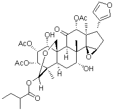
This is not surprising since Golgi and ER, which get disrupted during cell lysis, contain high amounts of both complete and partly synthesized glycans and these can alter the proportion of a particular glycan group. Each of the two glycan release methods was repeated six times to estimate their reproducibility. For nearly all glycan peaks, we observed higher coefficients of variation when glycome was analyzed from cell lysates. Especially high experimental variation was observed in highly branched glycan structures, which are important for the regulation of membrane half-life of many receptors. This increased variation of complex structures from experiment to experiment probably reflects their small contribution to the total glycome. Since glycans function as regulators of the activity of membrane proteins, many of them are also attached to proteins on the luminal side of various cytoplasmic vesicles, including the Golgi apparatus where posttranslational glycan processing occurs. Homogenization of cells results in mixing of these two compartments, containing physiologically separate fractions of glycans, and consequently leads to masking of relatively subtle, but functionally important, differences in the cell membrane Nglycome. Therefore, we believe that the analysis of Nglycome from embedded cells results in improved analytical precision due to elimination of the homogenization step from the procedure. Based on these observations we decided to further exploit the method of glycan analysis from embedded cells. We validated this hypothesis by mannosidase treatment, which resulted in Tubulin Acetylation Inducer almost complete disappearance of the corresponding chromatographic peaks, thus confirming the contribution of free oligomannose glycans to the pool of glycans released by PNGase F from glycoproteins associated with the cell membrane.
Category Archives: clinical trials
As cellular protein levels are not only dictated by capping efficiency where all variables unrelated to capping were constant
Cells originating from the same population were trasfected in order to over-express either the active HCE-WT-HA, the GTase-defective HCE-K294A-HA mutant, the GFP control protein or no protein. They were submitted to concentrations of 0 mM, 40 mM or 120 mM of mizoribine. All cell lines treated with mizoribine showed a global reduction in reporter protein expression when compared with untreated cells. This expected effect is likely due to partial guanosine pool depletion induced by IMPDH inhibition. Interestingly, the reduction in transcription and translation of the reporter was significantly less severe only in cells over-expression HCE-WT-HA for both mizoribine concentrations. The ability of HCE-WT-HA over-expression to partially rescue the luciferase expression in the presence of mizoribine ABT-199 demonstrates that HCE is one of the mizoribine pharmacological targets. Furthermore, the inability of the GTase defective mutant HCE-K294A-HA to rescue the reporter expression under mizoribine treatment further demonstrates, in agreement with our in vitro results, that in a cellular context it is the GTase activity of HCE that is targeted by MZP. Although indirect, this is strong evidence that mizoribine is able to impair capping in a cellular environment. As expected, capping could not be fully inhibited in cellulo at mizoribine concentrations of 40�C120 mM, which is approximately the in vitro IC50 of 80 mM. Despite its oral bioavailability and its low binding to serum proteins, mizoribine was not expected to reach intracellular concentration higher then its IC50. Of notice, the effect of mizoribine on cellular capping was however greater than anticipated. We hypothesize that the competition between HCE and Xrn2 for the nascent mRNA 59 end could explain the potency of MZP in cellulo, as slowing the capping activity could be sufficient to shift the balance towards quality control take-over and reduce downstream protein expression. What is the exact contribution of the capping apparatus inhibition to the global mizoribine mechanism of action? This question has yet to be addressed, but the immunosuppressive effect of mizoribine on T-cell is mainly mediated by GTP depletion and might be exacerbated by the reduction of mRNA capping and downstream cap-dependent translation. Our study clearly demonstrates that the therapeutic agent mizoribine monophosphate SB431542 301836-41-9 inhibits the human RNA guanylyltransferase in vitro and impairs mRNA capping in cellulo. Estrogen receptor negative breast cancer types are generally more aggressive and prone to metastasize. The absence of Estrogen receptor-alpha correlates with hormone-independent growth of these mammary tumor cells and causes uncontrolled proliferation and insensitivity to anti-hormonal treatments. In ERa-negative cell lines, a subset of genes is epigenetically silenced, while the majority of genes involved in cell cycle control and proliferation are constitutively expressed. Aberrant gene expression is frequently the result of chromatin modifications and composition, including histone posttranslational modifications and/or incorporation of histone variants. In particular, deregulation of enzyme complexes responsible for histone acetylation and deacetylation can be associated with breast cancer progression and an increase in tumor malignancy. Thus, compounds that change chromatin modifications are a promising anti-cancer approach.
The available data do not show any correlation between genetic aberrations of PI3K/mTOR pathways and sensitivity of BEZ235
In addition to inhibiting cell cycle progression, BEZ235 caused apoptosis in two of six cell lines. The inhibition of cell cycle Compound Library progression is a known effect of BEZ235, even at lower doses. However, apoptosis appears in only some cancer cell lines and is more apparent at higher doses of BEZ235. BEZ235 efficiently inhibited mTORC1, a molecule AB1010 controlling both cell cycle and apoptosis that might lead to cell cycle arrest and apoptosis in KAT4C and KAT18. Understanding the mechanisms through which BEZ235 contributes to apoptotic cell death will require further study. ATC is by far the most aggressive of the four major histologic types of thyroid cancer. Chemotherapy has been applied to treat patients with ATC with response rates around 20 to 50%. Novel strategies to improve outcomes are needed. Among three combination therapy regimens, BEZ235 combined with paclitaxel had the best synergistic effect in four ATC cell lines. Cancer cells with activation of PI3K/mTOR signaling are more resistant to paclitaxel, and the co-administration of PI3K/mTOR inhibitors with paclitaxel improves therapeutic effects. This finding is of clinical relevance since paclitaxel, a microtubule stabilizer, has shown to achieve a 53% response rate in patients with ATC in a phase II clinical trial. The combination of BEZ235 with a microtubule depolymerizing drug vincristine also revealed promising effect in the treatment of sarcoma. Our data showed that the combinational effects of BEZ235 with inhibitors of DNA topoisomerase type I or type II in treating ATC were largely antagonistic. Similar antagonistic effects of inhibition both topoisomerase activity and PI3K/AKT pathway have been observed in ovarian cancer cells. Cells in S phase are more susceptible to topoisomearse inhibitors. BEZ235-induced accumulation of cells at G0/G1 phase may therefore reduce the therapeutic advantage of concurrent therapy with topoisomerase inhibitors, explaining the unfavorable combination effects of BEZ235 with irinotecan and etoposide. Daily treatment of BEZ235 significant retarded 8505C xenograft tumor growth during the therapeutic period. The inhibitory effect was less prominent after the discontinuation of therapy, suggesting that prolonged treatment may be necessary to maintain therapeutic efficacy. BEZ235 significantly degraded caspase-3 in 8505C xenograft tumors, indicating this compound may induce apoptosis in vivo. After discontinuation of BEZ235, the volume of 8505C xenograft tumors subsequently increased. This enlargement of tumor following cessation of therapy might be due to normalization of cell size and resumption of cell proliferation, as mTOR and its downstream proteins S6 Kinase 1 and 4E-BP1 play pivotal roles in this respect. No significant weight loss or illness was observed during study period, suggesting that this therapy may have a promising safety profile. A prior report demonstrated that the presence of genetic alterations of PTEN, PIK3CA and AKT1 correlated well with sensitivity to an AKT inhibitor, but had weaker correlations with an mTOR inhibitor. This discrepency suggests that mTOR activity does not depend solely on PI3K/AKT activity. The reported data of genetic alterations in 8 thyroid cancer cell lines was summarized. Limited genetic changes on RAS/RAF/ERK and PI3K/mTOR pathways were identified.
In order to evaluate if MZP could impair in cellulo capping we monitored its indirect impact on the translation of the firefly luciferase
RTase and GTase domains since the abolition of the RTase domain EX 527 HDAC inhibitor reduces HCE MZP susceptibility. In order to gain additional details on the MZP main binding site, molecular docking could be used. Unfortunately, the structure of both the RTase and the GTase domain are separately available, but the structure of the full-length protein is still not available. Nevertheless, we ran a molecular docking experiment of MZP on the GTase domain from HCE. The lack of RTase domain, which is predicted to participate in MZP binding, and the moderate flexibility of the N-terminal domain, which introduces a structural incertitude, does not allow for definitive conclusions to be reached but it is interesting to note that, in preliminary experiments, MZP favorably docks on the N-terminal region of the HCE GTase. Together, our results reveal that MZP inhibits the HCE GTase activity with a 5-to 25-fold specificity in comparison to other GTases. Although more work is yet required to confirm our hypothesis, these results raise the possibility that the GTase inhibition could be mediated by a conformational change hindrance upon binding of MZP to an allosteric binding site that is speculated to reside near the RTase-GTase inter-domain. Nevertheless, mizoribine is one of the first compounds to demonstrate a certain degree of specificity toward a single GTase, despite the high degree of conservation of this crucial family of enzyme. MZP displays a higher in vitro inhibition potency for the GTase reaction in comparison to the complete RNA capping reaction. This may simply be due to our experimental conditions where the RTase activity of HCE, which is partially inhibited by free Mg2+, becomes the rate-limiting step. However, in cellulo the RTase harbors a higher BEZ235 turnover rate than the GTase, which catalyzes the limiting step in RNA capping. In a cellular context we expect the efficiency of MZP to be dictated solely by its interaction with the GTase. Historically, very few GTase inhibitors have been developed, neither as scientific tools nor as therapeutic agents. More recently however, novel GTase inhibitors have been discovered. They include the allosteric inhibitor mycophenolic acid, the pyrophosphate analog foscarnet which acts as a product inhibitor, and ribavirin triphosphate, a GTP analog that is transferred to acceptor RNAs by GTase, leading to stable but inefficiently translated pseudo-capped RNA. The current study identifies MZP as a novel allosteric GTase inhibitor, which is speculated to block a crucial conformational change. The GTase activity being the ratelimiting step of the essential capping apparatus, all these GTase inhibitors are promising lead candidates for the development of novel selective capping inhibitors and lead the way to a new class of anti-cancer, antifungal, and antiviral drugs. What is the biological relevance of the present finding? Numerous studies have demonstrated the potency of MZP to inhibit the cellular IMPDH and to lower the intracellular guanosine nucleotide pool thereby limiting cell growth, but none have addressed its impact on the capping apparatus. Monitoring the capping efficiency in living cells is a great challenge as the cellular quality control machinery degrades unsuccessfully capped mRNAs. Since proper capping is crucial for mRNA transcription, export, stability and translation, it is possible to monitor the capping efficiency based on the translation of a reporter protein.
Mitochondrial division either genetically or pharmacologically with the mitochondrial division inhibitor
To inhibit cell death in models of ceramide-induced toxicity and myocardial ischaemia and reperfusion injury. Furthermore, it was recently shown that treatment with mdivi-1 protected against pressure induced heart failure by ameliorating left ventricular dysfunction and promoting angiogenesis. Increase in apoptosis and abnormal mitophagy noticed in pressure overload samples were also prevented when treated with mdivi-1. Cancers are likely to develop in the later stages of life, when the chances of developing heart diseases are equally high. Patients with pre-existing heart diseases are usually excluded or underrepresented in clinical trials, which aim to identify the efficacy and potential adverse effects of drugs. We have recently shown that doxorubicin administration at reperfusion exacerbates ischaemia reperfusion injury, which was prevented when co-administered with cyclosporin A. It is therefore necessary to investigate the off-target effects of anti-cancer therapeutics or adjunct therapies in stressed or diseased conditions such as ischaemia and reperfusion injury. Given that doxorubicin-induced cardiotoxicity may be mediated by an imbalance in mitochondrial fusion and fission, we investigated the effects mdivi-1 on doxorubicin-induced cardiotoxicity using the Langendorff model in na?ve and in conditions of ischaemia and reperfusion injury. A model of oxidative stress was used to record the time taken to depolarisation and hypercontracture of cardiac myocytes upon drug treatment and western blot analysis was used to PF-4217903 evaluate the levels signalling proteins. Data on the effects of mdivi-1 on the cytotoxicity of doxorubicin was also assessed in HL60 cell line. Doxorubicin treatment is known to cause cardiovascular toxicity due to the generation of reactive oxygen species and calcium overload. Previous research has demonstrated that doxorubicin induced toxicity affects mitochondrial bioenergetics and causes mitochondrial fragmentation. We demonstrate that doxorubicin induced dysfunction on the haemodynamic parameters of the hearts are reversed by mdivi-1, a relatively specific inhibitor of mitochondrial division. Doxorubicin induced effects of cardiac function has been reported in in vivo and in vitro studies. Doxorubicin has previously been found to reduce both left ventricular developed pressure and heart rate, also shown in this study. Interestingly, the presented data show that doxorubicin treatment in the na?ve hearts caused a drop in the heart rate readings as opposed to its effects in conditions of ischaemia and reperfusion injury where no significant decrease in the heart rate values were recorded. One possible explanation for this effect could be the level of oxygen, previously published work has indicated that doxorubicin-induced decrease in the heart rate was more prominent when the heart were perfused with 95% oxygen as compared to 20% oxygen. We also show that co-treatment with mdivi-1 abrogated the detrimental effects of doxorubicin on left ventricular developed pressure. Interestingly, treatment with mdivi-1 was shown to ameliorate left ventricular dysfunction caused by pressure overload heart failure as assessed by left ventricular chamber diameter and fractional shortening. Mitochondrial fragmentation is proposed to be a major player in exacerbation of heart failure, inhibition of fragmentation is therefore thought to confer cardioprotection. Recent research has indicated that mitochondrial dynamics play a crucial role in cell physiology and growing evidence suggests that a balance between mitochondrial fission and fusion plays a vital role in pathological conditions. Studies have also shown that mitochondrial oxidative stress, which is also induced by doxorubicin treatment, leads to fragmentation of the mitochondria, which were attenuated with reactive oxygen species scavengers. Mitochondrial fragmentation has been found to mediate cellular function and apoptosis. Mdivi-1 has been suggested to have therapeutic potential for a variety of diseases such as stroke, myocardial GSI-IX side effects infarction and neurodegenerative disorders. In the current study, flow cytometric analyses of p-Drp1 levels show a significant up regulation of p-Drp1 levels following treatment with doxorubicin, which was prevented when doxorubicin was co-administered with mdivi-1. Elevated levels of mitochondrial fission proteins have been reported in response to ceramide and doxorubicin induced toxicity. It has been demonstrated that mdivi-1 inhibits GTPase activity by blocking self-assembly of Drp1.